

Diagnosis Reveals Tapeworm Larvae Cysts in Brain After Months of Hallucinations
A patient suffering from persistent headaches and visual disturbances was diagnosed with neurocysticercosis, a parasitic brain infection caused by pork tapeworm larvae.
By The Global Wire Newsroom · Reported from Rachel Melegrito
Link preview · horizonglobalnews.com
Diagnosis Reveals Tapeworm Larvae Cysts in Brain After Months of Hallucinations
A patient suffering from persistent headaches and visual disturbances was diagnosed with neurocysticercosis, a parasitic brain infection caused by pork tapeworm larvae.
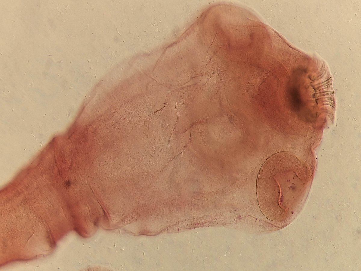
A medical case involving a woman who suffered from persistent headaches and visual hallucinations for several months culminated in a diagnosis of parasitic brain cysts containing tapeworm larvae, according to reporting published on Sept. 14, 2026, by medical journalist Rachel Melegrito. The patient's symptoms, which initially baffled clinicians during months of neurological disturbance, were ultimately linked to a parasitic infection of the central nervous system. The condition, known medically as neurocysticercosis, occurs when larvae of the pork tapeworm establish fluid-filled cysts within brain tissue, triggering localized inflammation, increased intracranial pressure, and severe neurological impairment.
Key facts
What happened
According to reporting by Rachel Melegrito, the patient spent months seeking medical answers while suffering from recurrent, severe headaches alongside visual hallucinations. Unexplained hallucinations combined with persistent cranial pain present significant diagnostic hurdles, as these symptoms can mimic psychiatric disorders, migraines, brain tumors, or vascular abnormalities.
After months of unresolved illness, comprehensive medical evaluations—typically involving neuroimaging technologies—uncovered fluid-filled cysts in the patient's brain. Clinical examination identified these lesions as housing tapeworm larvae, confirming an intra-cranial parasitic infestation.
While the published report by Melegrito highlights the patient's protracted timeline of symptoms and the dramatic eventual diagnosis, the specific clinical setting, hospital location, and precise diagnostic imaging details were not detailed in the original reporting. In standard clinical practice, diagnosing central nervous system parasitic cysts relies on Computed Tomography (CT) or Magnetic Resonance Imaging (MRI) scans, which can visualize individual larval scoleces—the anterior attachment structures of the tapeworms—within cystic lesions inside brain tissue.
Why it matters
The diagnostic discovery of brain tapeworm larvae underscores the diagnostic complexity of parasitic central nervous system infections and highlights broader public health vulnerabilities regarding food safety and hygiene. Neurocysticercosis is recognized by the World Health Organization (WHO) as a major neglected tropical disease, yet its diagnostic delay remains widespread even in modern health systems. When patients present with psychiatric or sensory symptoms such as visual hallucinations alongside chronic headaches, parasitic etiology is frequently overlooked in initial differential diagnoses, leading to months of unnecessary pain and risk of permanent neurological damage.
From a clinical and epidemiological perspective, the case illustrates the serious consequences of Taenia solium transmission pathways. Unlike intestinal tapeworm infections caused by eating undercooked pork containing larval cysts, human neurocysticercosis occurs when individuals inadvertently ingest microscopic tapeworm eggs shed in the feces of an infected human carrier. This can happen through contaminated food, water, or unwashed hands, making the disease a key indicator of sanitation standards and agricultural hygiene.
Furthermore, severe central nervous system infestations carry substantial healthcare risks. As larvae mature or die within brain tissue, the human immune system mounts a vigorous inflammatory response around the cyst walls. This localized swelling can lead to increased intracranial pressure, hydrocephalus, focal neurological deficits, and life-threatening status epilepticus. By shedding light on the presenting symptoms of headaches and hallucinations, cases like this reinforce the necessity for medical professionals to consider parasitic infections when evaluating unexplained sensory disturbances and chronic cranial distress.
The background
To understand how tapeworm larvae enter the human brain, it is necessary to examine the complex life cycle of Taenia solium, commonly known as the pork tapeworm. In a typical biological cycle, humans serve as the definitive host, harboring adult tapeworms in their small intestines after eating raw or undercooked pork containing viable larval cysts (cysticerci). These intestinal tapeworms produce thousands of microscopic eggs, which are excreted in human stool.
In contrast, neurocysticercosis develops when a human accidentally becomes an intermediate host by ingesting the parasite's eggs rather than the larvae. Once ingested, stomach acids dissolve the egg shells, releasing microscopic embryos called oncospheres. These embryos penetrate the intestinal wall, enter the bloodstream, and migrate to body tissues with high blood flow, notably the central nervous system, skeletal muscles, and eyes. Within brain tissue, the embryos develop into fluid-filled cysts containing invaginated tapeworm scoleces.
Globally, neurocysticercosis represents a major public health burden. According to epidemiological data from the World Health Organization, Taenia solium infection is responsible for an estimated 30 percent of all epilepsy cases in regions where the parasite is endemic, including parts of Latin America, Sub-Saharan Africa, South Asia, and Southeast Asia. In the United States, the Centers for Disease Control and Prevention (CDC) classifies neurocysticercosis as a Neglected Parasitic Infection, estimating that approximately 1,000 to 2,000 cases are hospitalized nationwide each year, predominantly among individuals who have lived in or traveled to endemic rural areas.
Standard medical management of neurocysticercosis depends on the location, stage, and number of cysts. As established in clinical literature from the Infectious Diseases Society of America (IDSA) and the American Society of Tropical Medicine and Hygiene (ASTMH), treatment often involves antiparasitic medications such as albendazole, sometimes combined with praziquantel, to kill the viable larvae. However, destroying intra-cranial cysts can trigger intense localized cellular inflammation as larval proteins are released into surrounding brain tissue. Consequently, physicians routinely administer high-dose corticosteroids, such as dexamethasone or prednisone, prior to or alongside antiparasitic drugs to prevent severe cerebral edema. In cases where cysts block the flow of cerebrospinal fluid, surgical intervention or ventriculoperitoneal shunt placement may be required.
Reaction
Following the publication of the case details by Rachel Melegrito, infectious disease specialists and public health experts routinely point to such clinical outcomes as reminders of the critical need for diagnostic vigilance. While specific public comments from treating physicians or the patient's family were not included in the original summary, medical professionals regularly emphasize in academic literature that visual hallucinations are a well-documented, though less common, manifestation of parasitic brain lesions.
Public health organizations, including the WHO and CDC, continue to advocate for integrated control programs aimed at eradicating Taenia solium. These initiatives focus on improved pig management practices, mandatory meat inspection, widespread community sanitation, personal hand hygiene education, and targeted mass drug administration in high-transmission zones.
What we don't know yet
Several critical clinical details remain undisclosed in the available reporting by Rachel Melegrito. The original report does not specify the age, geographic location, or travel history of the patient, leaving it unclear where and how the parasite was originally acquired. Geographic context is particularly vital for epidemiologists assessing whether a case represents local transmission or an imported infection from an endemic region.
Additionally, the reporting does not disclose the exact number, size, or anatomical placement of the tapeworm cysts within the patient's brain, nor does it specify the precise medical regimen or surgical interventions utilized during her care. The long-term neurological recovery of the patient—specifically whether her visual hallucinations and headaches completely resolved post-treatment—also remains unconfirmed.
What to watch
In the aftermath of this diagnostic case, public health officials and medical researchers will focus on several key indicators to assess transmission risks and clinical practices:
This report is based on original news reporting by Rachel Melegrito published on Sept. 14, 2026.
How this story was produced
This report was written by The Global Wire newsroom from reporting first published by Rachel Melegrito. We verify the core facts against the original report, write our own account, and add the background and consequences a short wire item leaves out. Drafting is AI-assisted inside an editor-supervised pipeline, and every story is checked for accuracy of attribution, structure and duplication before it appears — full detail in our AI and funding disclosure.
Spotted an error? Tell us at corrections@horizonglobalnews.com and read our corrections policy or editorial standards.



Reader comments
Loading comments…