

Skull Bone Marrow Immune Channels Linked to Chronic Head Pain in Neuroinflammation Study
Research into direct vascular pathways between cranial bone marrow and the meninges reveals how localized immune responses inside the skull may drive persistent neurological pain.
By The Global Wire Newsroom · Reported from Peter Dockrill
Link preview · horizonglobalnews.com
Skull Bone Marrow Immune Channels Linked to Chronic Head Pain in Neuroinflammation Study
Research into direct vascular pathways between cranial bone marrow and the meninges reveals how localized immune responses inside the skull may drive persistent neurological pain.
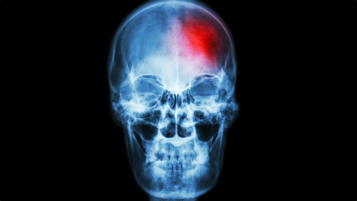
For decades, neuroscientists and immunologists operated under a foundational premise: when the brain encountered injury, infection, or localized tissue stress, the immune cells deployed to protect central nervous system tissues traveled from distant reservoirs in the body, such as the marrow of long bones in the arms and legs, circulating through the general bloodstream to reach the head. However, research into cranial anatomy and neuroimmunology is dismantling this long-held understanding. According to reporting by Peter Dockrill published on September 2, 2026, scientific investigation has revealed that specialized immune cells produced directly within the bone marrow of the skull play a far more direct, localized, and previously underappreciated role in central nervous system immune responses. Rather than relying solely on systemic circulation, the skull contains its own active marrow compartments that communicate directly with the membranes wrapping the brain. This localized immune infrastructure, while vital for rapid defense, is now emerging as a potential hidden contributor to persistent neuroinflammation and chronic pain conditions inside the head.
Key facts
What happened
The traditional model of neuroimmunology framed the central nervous system as an isolated region that relied on systemic immune responses during emergencies. Under this classical framework, hematopoietic stem cells in the bone marrow of large bones—such as the femur, tibia, or sternum—produced white blood cells including neutrophils, monocytes, and lymphocytes. When inflammation or injury occurred in the cranial cavity, these cells were thought to be transported through the peripheral arterial system, eventually crossing or interacting with the blood-brain barrier and meningeal blood vessels.
Recent scientific investigations detailed by Dockrill challenge this traditional narrative by uncovering a direct structural and functional bridge between the skull's internal marrow and the brain's outermost membrane layer. Within the spongy tissue layer of cranial flat bones—known as the diploë—active bone marrow maintains a local reservoir of immune progenitor cells. Microscopic anatomical studies have identified tiny vascular channels, or micro-vessels, that pass directly through the inner table of the skull bone, connecting the cranial bone marrow directly to the dura mater.
When tissue stress, trauma, or localized chemical signals occur in or around the brain, immune cells stored in the skull marrow do not enter general systemic circulation. Instead, they migrate directly through these microscopic conduits into the meninges. While this localized architecture enables rapid protection against localized infections or physical injury, researchers are discovering that persistent, dysregulated activity in these cranial marrow niches can lead to continuous immune cell discharge. This chronic influx of inflammatory cells into the meningeal space triggers localized tissue swelling and releases pro-inflammatory signaling molecules, directly irritating sensory nerve fibers and providing a biological mechanism for persistent chronic pain inside the head.
Why it matters
The identification of localized cranial immune trafficking marks a significant shift in how researchers understand and treat chronic neurological pain and neuroinflammatory disorders. Historically, conditions such as refractory migraines, tension-type headaches, post-traumatic headache syndromes, and trigeminal neuropathic pain were treated primarily through systemic pharmacotherapy—ranging from nonsteroidal anti-inflammatory drugs and triptans to systemic immunosuppressants and targeted monoclonal antibodies. However, systemic therapies often carry broad side effects and may not achieve adequate therapeutic concentrations within specific localized microenvironments of the skull and meninges.
By identifying the skull's bone marrow as an active, localized source of inflammatory cells, researchers open new pathways for localized therapeutic intervention. Understanding that cranial marrow channels provide a direct conduit to the dura mater suggests that medical treatments could be developed to specifically target these local cellular reservoirs or block the signaling cascades within cranial vascular channels. Such targeted approaches could potentially prevent the localized influx of inflammatory cells without suppressing the body's overall systemic immune system.
Furthermore, this mechanism offers a fresh perspective on why certain chronic pain conditions persist long after an initial head injury or infection has resolved. If the bone marrow inside the skull remains in a heightened state of inflammatory readiness, it may continuously pump activated immune cells into the meninges, keeping pain receptors persistently sensitized. Addressing the root cause of chronic pain may therefore require therapies that reset or quiet the immune microenvironment within the cranial bones themselves.
The background
To fully grasp the significance of skull bone marrow immune dynamics, it is necessary to examine the history of neuroimmunology and the evolution of scientific thought regarding the central nervous system. For more than a century, the medical consensus held that the brain enjoyed complete "immune privilege." This concept, popularized in the mid-20th century, suggested that the brain was entirely secluded behind the blood-brain barrier—a dense layer of endothelial cells, astrocytes, and pericytes lining cerebral capillaries—preventing circulating immune cells and peripheral pathogens from entering delicate brain tissue.
This paradigm began to undergo substantial revisions over the past decade. In 2015, structural discoveries identified functional lymphatic vessels lining the dura mater, proving that the central nervous system possesses a direct pathway for fluid drainage and immune cell trafficking to deep cervical lymph nodes. Shortly thereafter, in 2018, groundbreaking imaging studies in rodent models and human tissue specimens revealed the existence of microscopic, direct vascular channels connecting the bone marrow of the skull to the dura mater.
The skull bone consists of an outer compact bone layer, an inner compact bone layer, and a middle layer of cancellous, spongy bone known as the diploë. The diploë contains active red bone marrow throughout adult life, capable of hematopoiesis—the production of cellular blood components. The dura mater, the outermost of the three meningeal layers covering the brain, is richly innervated by sensory nerve fibers from the trigeminal nerve, designated as cranial nerve V. Trigeminal nerve endings in the dura are primary pain-sensing structures in the head; when exposed to inflammatory cytokines, histamine, or calcitonin gene-related peptide released by activated immune cells, these nerve fibers fire continuous pain signals to the brainstem.
Historically, researchers assumed that all bone marrow compartments in the human body functioned identically, responding uniformly to systemic endocrine and cytokine signals. However, recent comparative studies between cranial marrow and long bone marrow have revealed key molecular differences. Skull marrow exhibits distinct gene expression profiles, specialized stromal cell populations, and a unique sensitivity to local central nervous system cues, establishing it as a functionally distinct immune niche tailored specifically for cranial protection—and, when dysregulated, cranial pathology.
Reaction
The neuroscientific and pain research communities have responded to these localized immune discoveries with widespread interest, as clinicians and basic researchers re-examine long-standing clinical puzzles. Pain specialists have noted that localized cranial immune activity provides a plausible biological mechanism for treatment-resistant headache syndromes that fail to respond to standard vascular or neuro-targeted drugs.
Neuroimmunologists are currently calling for expanded translational research to map the precise cellular communication between cranial bone marrow and the meninges in human patients. Pharmacologists and drug developers are evaluating how future therapeutic compounds might be delivered directly to cranial marrow spaces or designed to selectively modulate the micro-channel transport systems connecting the skull to the dura.
Because Dockrill's reporting focuses on basic scientific mechanisms and ongoing discoveries regarding skull immune pathways, formal clinical position statements from major neurological societies remain forthcoming. However, consensus among researchers in the field indicates that future diagnostic criteria for neuroinflammatory and chronic pain conditions will increasingly incorporate localized bone marrow assessments alongside traditional brain imaging and cerebrospinal fluid analysis.
What we don't know yet
Despite significant advances in characterizing the physical channels and cellular migration between the skull marrow and the meninges, critical questions remain unanswered. First, researchers have yet to determine the precise molecular signals that trigger skull bone marrow to initiate or sustain hyperactive immune cell production in patients suffering from chronic pain. It is unclear whether local neural signals, circulating chemical messengers, or physical mechanical stress on the skull are the primary drivers of this dysregulation.
Second, the degree to which cranial marrow architecture and cellular output vary across different human populations remains uncertain. Factors such as age, biological sex, genetic predispositions, and previous history of traumatic brain injury or viral infections may significantly alter the density of cranial vascular channels or the baseline activity of skull marrow.
Finally, researchers do not yet know how to selectively modulate skull bone marrow function without compromising necessary local immune defenses. Because these cells serve a vital role in protecting the brain against ascending pathogens and promoting tissue repair after injury, completely shutting down localized immune output could leave the central nervous system vulnerable to severe infections. Developing therapies that specifically target chronic pain pathways while preserving protective immunity represents a major ongoing challenge.
What to watch
In the coming years, several key developments will indicate how this research translates into clinical practice and therapeutic innovation. Researchers will be monitoring ongoing advanced imaging studies utilizing high-resolution micro-computed tomography and high-field magnetic resonance imaging designed to map the density and structural variations of cranial vascular channels in living human patients.
Watch for upcoming clinical studies investigating cellular biomarkers in individuals with chronic pain conditions such as persistent post-traumatic headache, cluster headache, and trigeminal neuralgia. Identifying specific inflammatory cell profiles originating from cranial marrow could lead to novel diagnostic tools and blood or tissue tests.
Additionally, pharmaceutical researchers are expected to initiate preclinical trials testing localized drug delivery systems, such as targeted topical or targeted micro-vascular therapies, designed to reach cranial bone marrow niches directly. Results from these early-stage therapeutic trials will determine whether modulating skull-specific immune pathways can successfully alleviate chronic cranial pain in clinical settings.
This article is based on reporting originally published by Peter Dockrill for ScienceAlert on September 2, 2026.
How this story was produced
This report was written by The Global Wire newsroom from reporting first published by Peter Dockrill. We verify the core facts against the original report, write our own account, and add the background and consequences a short wire item leaves out. Drafting is AI-assisted inside an editor-supervised pipeline, and every story is checked for accuracy of attribution, structure and duplication before it appears — full detail in our AI and funding disclosure.
Spotted an error? Tell us at corrections@horizonglobalnews.com and read our corrections policy or editorial standards.





Reader comments
Loading comments…